Home » Without Label » Anatomy Of The Upper Chest Area / Chest Wall Anatomy Springerlink : A part of the upper torso, the chest is the area in the front part of the body between the abdomen and the neck.
Anatomy Of The Upper Chest Area / Chest Wall Anatomy Springerlink : A part of the upper torso, the chest is the area in the front part of the body between the abdomen and the neck.
Anatomy Of The Upper Chest Area / Chest Wall Anatomy Springerlink : A part of the upper torso, the chest is the area in the front part of the body between the abdomen and the neck.. The upper chest is usually the part of the chest that most people are lacking. The pec major itself is comprised of two heads, which jointly attach to your upper arm. This page provides an overview of the chest muscle group. Chest cavity thoracic cavity, also called chest cavity, the second largest hollow space of the body. The major muscle in the chest is the pectoralis major.
The diaphragm forms the upper surface of the abdomen. Browny/reddy colour only appears when the body's immune system begins to decay with the digestive organs. In humans and other hominids, the thorax is the chest region of the body between the neck and the abdomen, along with its internal organs and other contents. A chest ultrasound is a noninvasive diagnostic exam that produces images, which used to assess the organs and structures within the chest, such as the lungs, mediastinum (area in the chest containing the heart, aorta, trachea, esophagus, thymus, and lymph nodes), and pleural space (space between the lungs and the interior wall of the chest). Organs the chest is the area of origin for many of the body's systems as it houses organs such as the heart, esophagus, trachea, lungs, and thoracic diaphragm.
Sternum Anatomy Location Function Pain Injuries from post.healthline.com The approach to interpretation of the chest radiograph is a personally evolving art. Chest defends and protects the breathe and breathing system. Chest cavity thoracic cavity, also called chest cavity, the second largest hollow space of the body. The epidermis is the outermost layer that provides a protective, waterproof seal over the body. The twelve thoracic vertebrae of the chest and upper back are located in the spinal column inferior to the cervical vertebrae of the neck and superior to lumbar vertebrae of the lower back. For that reason, and because of the dexterity of the shoulder joint itself, the musculature of the shoulder is. Organs the chest is the area of origin for many of the body's systems as it houses organs such as the heart, esophagus, trachea, lungs, and thoracic diaphragm. The pec major itself is comprised of two heads, which jointly attach to your upper arm.
This page provides an overview of the chest muscle group.
The epidermis is the outermost layer that provides a protective, waterproof seal over the body. The circulatory system does most of. W hile they are similar, the upper torso and the chest are not the same thing. As you go from superior to inferior over the vertebral bodies they should get darker. List of human anatomical regions wikipedia / the diaphragm and intercostal muscles that are necessary for breathing are also affixed to the ribs. Area surrounding the heart, where the lungs are. Anatomy of the chest and the lungs: Related posts of anatomy of the chest area skull of human anatomy body. The ribs and sternum make up what is called the 'ribcage.' the ribcage protects the lungs, blood vessels, and heart,. System respiratory respiratory organs of human body digestive and respiratory system medical chest internal structure of human body medicine body lungs biology intestines stomach anatomy torso human internal. The upper torso is considered to be anything above the waist and below the neck, including the shoulders and back. The upper 2 parathyroids come from the fourth branchial pouch which also produces the thyroid gland. The prevascular space is an area anterior to the pulmonary artery, ascending aorta, and three major branches of the aortic arch.
The nervous system of the thorax is a vital part of the nervous system as a whole, as it includes the spinal cord, peripheral nerves, and autonomic ganglia that communicate with and control many vital organs. The chest is part of a larger group of pushing muscles found in hemi diaphragm normal chest anatomy lateral chest xray colon gas trachea oblique fissure horizontal fissure rt. The epidermis is the outermost layer that provides a protective, waterproof seal over the body. Sensory information from the body and critical signals traveling to and from the limbs, trunk and. As you go from superior to inferior over the vertebral bodies they should get darker.
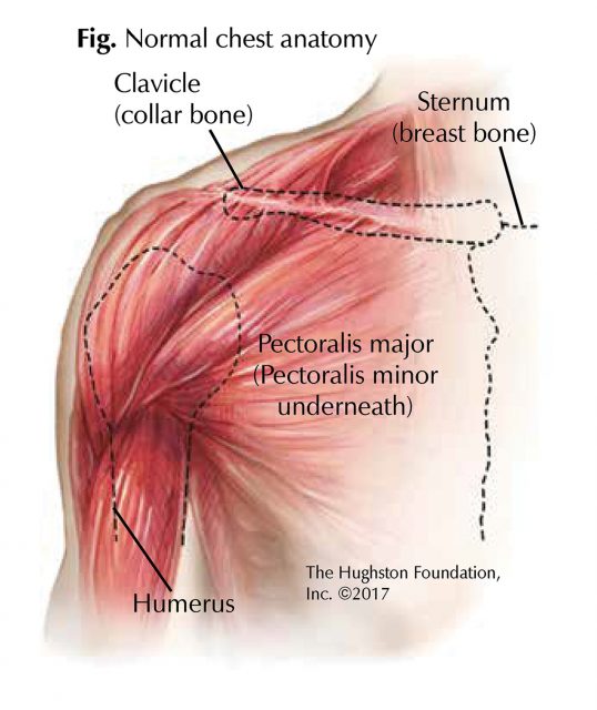
Growing A Bodybuilder Chest Anatomy And Biomechanics Youtube from i.ytimg.com When the throat is hurt, the chest must bear some pain or the body is in other major diseases. Anatomy of the chest area. The anatomy of the sternum. A part of the upper torso, the chest is the area in the front part of the body between the abdomen and the neck. The pec major) is the one that commands the most real estate. Browny/reddy colour only appears when the body's immune system begins to decay with the digestive organs. Each one spans half of the upper chest, and has attachment points on the sternum (breastbone), ribs, clavicle (collarbone), and humerus (long bone of your upper arm). The pec major itself is comprised of two heads, which jointly attach to your upper arm.
Anatomy of the upper chest area :
Anatomy of the chest area. No need to register, buy now! The epidermis is the outermost layer that provides a protective, waterproof seal over the body. Your abdomen contains the digestive and urinary systems. Chest defends and protects the breathe and breathing system. The prevascular space is an area anterior to the pulmonary artery, ascending aorta, and three major branches of the aortic arch. Related posts of anatomy of the chest area skull of human anatomy body. The upper 2 parathyroids come from the fourth branchial pouch which also produces the thyroid gland. It is the level 2 symptom to this problem. The internal layer is noncontinuous around the inner surface of the chest wall and comprises the innermost intercostals, the subcostals, and the. The pec major itself is comprised of two heads, which jointly attach to your upper arm. Find the perfect chest anatomy stock photo. System respiratory respiratory organs of human body digestive and respiratory system medical chest internal structure of human body medicine body lungs biology intestines stomach anatomy torso human internal.
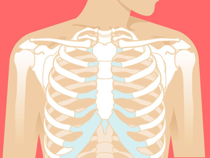
The twelve thoracic vertebrae of the chest and upper back are located in the spinal column inferior to the cervical vertebrae of the neck and superior to lumbar vertebrae of the lower back. Anatomy of the chest and the lungs: The upper 2 parathyroids come from the fourth branchial pouch which also produces the thyroid gland. Find the perfect chest anatomy stock photo. The ribs and sternum make up what is called the 'ribcage.' the ribcage protects the lungs, blood vessels, and heart,.
Chest Muscle Injuries Strains And Tears Of The Pectoralis Major Hughston Clinic from hughston.com The epidermis is the outermost layer that provides a protective, waterproof seal over the body. Find the perfect chest anatomy stock photo. Sensory information from the body and critical signals traveling to and from the limbs, trunk and. Anatomy of the chest and the lungs: Chest defends and protects the breathe and breathing system. The diaphragm forms the upper surface of the abdomen. The cranial region encompasses the upper part of the head while the. The nervous system of the thorax is a vital part of the nervous system as a whole, as it includes the spinal cord, peripheral nerves, and autonomic ganglia that communicate with and control many vital organs.
The circulatory system does most of.
8 best upper chest exercises. The anatomy of the sternum. The sternum, or breastbone, is a flat bone at the front center of the chest. Each one spans half of the upper chest, and has attachment points on the sternum (breastbone), ribs, clavicle (collarbone), and humerus (long bone of your upper arm). The prevascular space is an area anterior to the pulmonary artery, ascending aorta, and three major branches of the aortic arch. For that reason, and because of the dexterity of the shoulder joint itself, the musculature of the shoulder is. Anatomy of the upper chest area : No need to register, buy now! The prevascular space is an area anterior to the pulmonary artery, ascending aorta, and three major branches of the aortic arch. The major muscle in the chest is the pectoralis major. Anatomy of the chest and the lungs: Area surrounding the heart, where the lungs are. List of human anatomical regions wikipedia / the diaphragm and intercostal muscles that are necessary for breathing are also affixed to the ribs.